IANSA - Interdisciplinaria Archaeologica Natural Sciences in Archaeology ● Volume I ● Issue 1–2/2010 ● Pages 77–89
News and views
Congenital Cranial Defect in a Female from a Funnel Beaker Culture Settlement Burial in Náměšť na Hané (Czech Republic) – a Case Report
Anna Pankowská*, Jaroslav Peška, Jakub Vrána
Archaeological Centre, Archeologické centrum Olomouc U Hradiska 42/6, 779 00 Olomouc, Czech Republic
*Corresponding author. E-mail: annapankowska@gmail. com
Abstract
Scaphocephaly, a congenital defect of the skull, was identified in an adult female skeleton interred in a Funnel Beaker culture settlement pit (4590±30 years BP) in central Moravia. Scaphocephaly is a natural deformation of the skull resulting from premature fusion of sagittal sutures. Premature fusion (craniosynostoses) can cause deformation of the skull (craniostenoses). The skull in question is deformed and disproportionately long and narrow (cranial index: 55). Fusion probably occurred in early childhood, most likely shortly after birth, when the brain was growing most quickly. Scaphocephaly is usually not associated with mental or physical disorders, however, the greater the deformation the greater the chance of intracranial pressure and compression of the brain. Some studies suggest that non-syndromic single suture fusion influenced intellectual development in half of examined cases. In clinical research individuals with scaphocephaly demonstrated asymmetric cerebral blood flow. The etiology of these defects is not fully understood. In addition to the congenital defect of the skull, there are Harris lines on the metaphysis of the tibiae. We used visual observation, metric analysis, CT scan and X-ray examination to precisely record all pathological lesions. These pathological findings are an important contribution to the archaeological context and circumstances of the funeral, and may be evidence of the treatment of deformed individuals. While this woman might not have been affected by a mental disorder, her appearance differed markedly from what was normal in her society. It is possible that the funeral practice related to her remains was a reflection of her social status. From an anthropological point of view this is a uniquely well-preserved human remain for the period, and is of local and world-wide importance.
Article info
Article history:
Received: 6 December 2010
Accepted: 2 February 2011
Key words:
Funnel Beaker culture
congenital anomaly
scaphocephaly
settlement burial
freshwater pearl mussel shells
1. Location and circumstances of the find
During the rescue excavation conducted in connection with the reconstruction of the Loučany to Náměšť na Hané road, Neolithic (Linear Pottery culture) and Eneolithic (Funnel Beaker culture) settlements were discovered in the centre of the town of Náměšť na Hané (Figure 1). Our attention was drawn to a human skeleton of feature No. 7 which was partially filled with soil. In this feature – an irregular oval shaped settlement pit – a pathological female skeleton was found with an unusual mother-of-pearl bead necklace (Vrána 2010; Vrána, Pankowská 2010).

Figure 1. Map of the town Náměšť na Hané, showing location of site with the skeletal remains examined in this study (www. mapy. cz).
The feature and skeleton are dated to the Funnel Beaker culture as are other features (Nos. 1, 6, 8) that intersect borrow pit No. 7 (Figure 2). M. Šmíd dates pottery from the pit to the Baalberg phase of the Funnel Beaker culture. The human female skeleton was found in an almost straight position (Figure 3) in the NW part of the area, between feature 6 and the border of feature 7. There is no evidence of a grave pit (Vrána, Pankowská 2010).
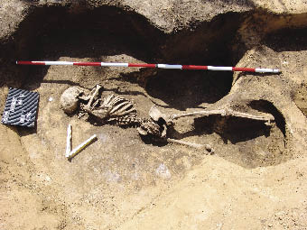
Figure 2. Photograph of the female skeleton interred in the borrow pit (photo by Anna Pankowská).

Figure 3. Plan of rescue excavation (plan by Pavel Grenar; drawing by Antonie Pešková).
1.1 Absolute dating
The discovery of several dozen small cylindrical mother-of-pearl beads (Figure 4) inside the mandible of the skeleton in question provoked discussion as to the dating of the whole site. Therefore a decision was made to take a sample of human bone (phalanx from the upper left extremity) for absolute dating. Analysis was performed at the University of Georgia (USA), with a result of UGMAS # 05712: 4590 ±30 bp (uncalibrated), which makes 1 sigma 3380–3340 BC or 2 sigma 3380–3330 BC after calibration (Figure 5). This date corresponds to the late Boleraz phase or the turning point to the classical Baden Culture in Austria, the late Lubel-Volyn culture, or late phases of southeastern groups of the Funnel Beaker culture in Poland (Stadler et al. 2001; Zakościelna 2009; Włodarczak 2006). The remains belong to a period later than any other published Eneolithic finds from Hlinsko (Pavelčík 1992, 2002). They precede the recently published findings from the necropolis of the classic Baden culture from Lower Austrian Ratzersdorf (Krumpel 2009). In any case this absolute date is later than the early period of the Funnel Beaker culture. Even the position of the skeleton in the partly filled pit (probably a clay pit from the Linear Pottery culture or early Funnel Beaker culture) may help us in placing the burial somewhere in the period of the Boleraz culture or the beginning of the classic Baden culture. While there is little Baden culture material remains evident at the site, this may be due to the restricted scale of the rescue excavation.
Although settlement burials are frequent in the Funnel Beaker culture (e. g. Zápotocký 2008) they can also be found in other prehistoric periods, from the Neolithic to Early modern period. They differ in quantity by particular ethnic groups. For some it is a standard type of burial, for others it is an exceptional phenomenon (Tichý in press).

Figure 4. Tiny beads of freshwater pearl mussel shells (Margaritifera margaritifera) (photo by Mojmír Bém).
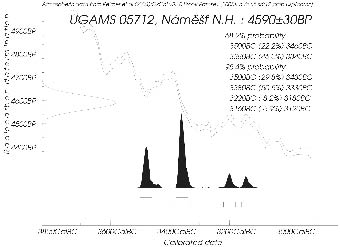
Figure 5. Graphical illustration of radiocarbon dating probabilities.
1.2 Comparable finds
A set of small beads shaped as flat discs with crosswise holes was found piled on the mandibular ramus of the deceased. These beads are very small (around 5 mm in diameter and about 2–3 mm in height), and according to M. Nývltová-Fišáková cut from the shells of pearl mussels. Their original number is indeterminate as most are fragmented. Even though we have evidence of mother-of-pearl beads, mainly from several necropoles of the Early Bronze Age we can also identify them in many prehistoric communities older and later than these remains from Náměšť na Hané. Older beads are slightly bigger (151 pieces) and made of different species of pelecypods, such as those found in the lumbar region of an adult female from a Linear Pottery culture necropolis in Samborzec 1 (grave III – obj. 208, Czekaj, Zastawny 2009). Less clear is an older finding of the Moravian Painted Ware culture in Džbánice, where a necklace made of shells was found on skeleton No. VIII, within a mass grave site. While Podborský mentions “beads cut out of shells of local pelecypods” (Podborský 1993), the original publication of the find (Horňanský, Skutil 1950) speaks only about dentalia or spondylus jewelry. In approximately the same period we notice a mass occurrence of beads made from Unio sp. fresh water shells in the Brześć Kujawski Lengyel culture. They occur in two different forms, as rectangular tablets (two or four holes and parts of necklaces), or as round beads (one hole and as parts of loin belts, necklaces, and sometimes in combination with shells from other species of pelecypods, animal teeth, copper beads or ornaments). They occur very frequently (around 8 thousand pieces) and are typical for this cultural group. They come exclusively from female graves at sites such as Brześć Kujawski 4, Osłonki 1 and Konary 1 (Grygiel 2004; 2008). The discovery of their manufacturing site in a neighboring settlement dated to the same period (Brześć Kujawski 4, pit 871), where a large accumulation of production waste was discovered (Grygiel 2008; 1866), is an important find. The mother-of-pearl beads formed parts of necklaces in the same culture also at the site of Kruszy Zamkowej (Bednarczyk, Czerniak, Kośko 1980).
They are also found within the lower (Wiślański 1979) and middle Eneolithic, specifically from the Funnel Beaker and Baden cultures. A higher occurrence of beads is also found in the Lubel-Volyn culture in SE Poland (Zakościelna 2009). It is interesting that grave No. 5. in the necropolis of Grodek IC is of the same date as the grave from Náměšť na Hané (Zakościelna 2009, Tab. VIII: 4570±70 BP). In the aforementioned group different types of beads and snail shells were also found as, for example, in 3 graves in Złota, “Grodzisku” I (Zakościelna 2009). Twenty-six beads made of mother-of-pearl (fresh water Unio sp. ) were found in the upland Baden culture settlement at Hlinsko near Lipník nad Bečvou. At this site necklaces composed of animal teeth, deer teeth, copper, limestone, bone and mother-of-pearl beads, completed by pendants made of mother-of-pearl, copper and terracotta were also found (Pavelčík 1989, 1993). With the exception of one piece from the upland settlement of Staré Zámky, in Brno-Líšeň, (Medunová-Benešová 1964) we can find parallels outside of the Czech Republic, such as the Baden culture necropolis in Hungary, where whole shells were found (Banner 1956). They are also common in the later Trypillian culture, where their frequency is so high that their use as currency is more likely than as decoration (Markevič 1981).
The climax of their prevalence in Central Europe occurs in the Corded Ware culture where they are found abundantly as parts of necklaces, patches on dresses, or on the hems of clothes in many different regions including Germany, Bohemia, Moravia and the region of “historic Lesser Poland” (south Poland) (Buchvaldek 1986, Włodarczak 2006). They are scarce in the related Złota culture (Machnik 1979) and in the latest, and surprising find, in SW Slovakia (Krakovany-Stráž), beads are found scattered all over the grave (Klčo, Krupa 2008). According to E. Neustupný (2008) they are typical for female graves of the Middle and Late Corded Ware culture often in combination with pendants made of animal teeth with holes in them. In one abundant find (1,453 pcs. of mother-of-pearl beads) in Prague-Čimice, at least 4 necklaces were found, with an additional small necklace of mother-of-pearl rings found under animal teeth at the waist level of the skeleton, the largest of which lay across the chest in 6 rows. Havel (1981) contemplates the possibility of the scattering of decorations over the body as part of the burial practices. A similar situation is observed in a find of 6,164 pieces of beads (L. Šebela indicates 5,464 whole beads and 1,700 fragments) concentrated under the back of the skeleton in a rich grave from Marefy (Chleborád 1934, Šebela 1999). Another Moravian example is the 73 beads from the graves found in Kyjov (Šebela 1999, 84). The presence of such beads in the Bell Beaker Culture is questionable as the grave at Sulejovice (cf. Moucha 1958; Neustupný, Smrž 1989) is dated to the Corded Ware culture. The grave complex from Most (uncertain context; Moucha 1958, 72) includes various objects characteristic of the Corded Ware culture (mother-of-pearl beads, drilled animal teeth and their imitations) and the Bell Beaker culture. The complex is interpreted as two graves, one the Corded Ware and one from the Bell Beaker culture, superimposed (Dobeš, Buchvaldek 1993). The rather rare find of mother-of-pearl beads in a Globular Amphora culture grave at Brozany nad Ohří in NW Bohemia is evidence that these beads are not chronological or cultural indicators (Dobeš 1998; 2008).
Decorations made of mother-of-pearl are found again at the beginning of the Bronze Age. Their origin is tied to a unique grave (# 2) under burial mound #14 in the Catacomb Culture burial ground in Vinogradno, where nacre and unfinished nacre products from the shells of Unio sp. (37 pcs.) were found. According to Bátora (2002), this child burial was protected by the magical power of mother-of-pearl beads. Good contribution as to the question of the origins of mother-of-pearl beads is also their great number found at the burial grounds of the Maroš/Mureş Culture in Mokrin (Girić 1971; Kadrow 1998, 257). In central Moravia they appear in Carpathian epi-Corded Ware assemblages, less-so in the Nitra culture, and more often in the Košťany, Mierzanowice and Strzyówska cultures. In the Košťany culture in particular, they can be found from the earliest period, and from the classic period there is evidence of local manufacture, from graves #101 in Košice and #54 in Košťany (Bátora 1982). Evidence of workshops is also known from the Mierzanowice culture in Iwanowice, Babiej Góry I (feature 48) or Babiej Góry II (feature 82 and 457) with appropriate inventory of graves (Kadrow, Machnikowie 1992). At this location they can be also found in the necropolis of late phase Szarbia, whereas in the Nitra culture area they are associated mainly with the classic period (Bátora 2000). Similar examples are found in graves at Komjatice, Výčapy-Opatovce, Jelšovce or Pobedim (Točík 1978, 1979; Bátora 2000; Bialeková 2000).
In Moravia, mother-of-pearl beads can be found in the burial ground at Holešov (6×), concentrated around the skull, knee joints, pelvis, chest and tibiae. Similar layout of approximately 1000 little beads was found in grave n. 11 at the burial ground in Slatinice (Šmíd 2006). The variety of bead placement in graves led Šmíd to conjecture that these beads were generally used for necklaces, hair decorations, tufts on clothing, as decorative patches or on belts, including exotic embroidery on skirts and shoes (Šmíd 2006).
No matter the normal variety in the placement of mother-of-pearl beads, in the case of Náměšť na Hané we can safely conclude that the beads were used as a neck decoration. After the decomposition of organic remains, including the strap and string, the beads settled in the mandible. That these beads are found alongside a rather poor set of grave inclusions corresponds with most previous findings. This brief overview clearly demonstrates these beads are not chronological or cultural indicators. However, the absolute date of the find from Náměšť na Hané and the absence of a contemporary cultural horizon in the area of the rescue excavation are surprising. The anomalous burial of the deceased female may be connected with her handicap, which became the subject of anthropological analysis.
2. The Skeleton
An adult female skeleton was interred in an anatomically correct position on its left side (SV–JZ). Certain skeletal parts were found at different levels within the pit fill since the individual had not been interred at the bottom of the borrow pit (Figure 2, 3). Joints were in close anatomical position, due to the borrow pit having been filled in shortly after the internment of the individual. From the position of the skeleton we cannot determine whether the individual was intentionally buried or simply tossed into the settlement pit. But the aspect of the upper right extremity cannot be considered a consequence of random disposal of the dead body. The intentional arrangement of the upper right limb and the rest of the body cannot be excluded. This internment is characterized by a soil covering deposited shortly after death, positioning of the limb, and placement of beads of freshwater pearl mussel shells inside the mandible. We can ascribe the latter to a burial, though we are unable to distinguish whether we are dealing with the ritual or non-ritual disposition of the individual. In contrast to the customary Funnel Beaker culture burial, this female was not correctly interred, but disposed of in a special manner – within a settlement pit.
3. Methods of demographic and paleopathological assessment
After field preparation, documentation, excavation and laboratory clearance, the quantitative preservation (%), stature, age and sex of the skeleton was estimated. Quantitative preservation was calculated according to Stojanowski et al. (2002), and stature was estimated according to Sjøvold (1990). Age was estimated on the basis of two methods, from the pubic symphyseal surface (Brooks, Suchey 1990), and from the auricular surface of the ilium (Lovejoy 1985). Determination of sex is based on measurements of the pelvis (Murail et al. 2005). The macroscopic assessment of cranial suture ossification was accomplished using Masset’s method (Masset 1989).
Examination of pathological traces was assessed first on a macroscopic level. Following which we measured the skull (following Knussman 1988; Drozdová 2004) using slide and spreading calipers. Cranial capacity was evaluated following the formula of Pearson (1935). X-ray and CT scan images were made in the Radiodiagnostic Department of the First Faculty of Medicine, Charles University in Prague. Finally, we compared the measurements with 4 scaphocephalic skulls from the collection of the 2nd Pathological Institute of the National Museum in Prague, three of which have a plaster cast of the brain and traces of intracranial pressure on the endocranium.
4. Results of demographic parameters and description of pathological lesions
The skeleton is very well preserved. According to the method of Stojanowski et al. (2002) 78 % of skeletal elements are present. We were able to collect all required osteological data. Skeletal elements are complete and undamaged. There is no evidence of postmortem destruction, mammal activity or traces of plant roots. The skeleton belongs to a female (Murail et al. 2005) 30 – 53 years old (Brooks, Suchey 1990) and 158–168 cm tall (Sjøvold 1990).
Table 1. Degree of synostosis (Masset 1989).
|
SUTURA |
Unilaterally |
DX |
SIN |
|
C1 |
– |
2 |
1 |
|
C2 |
– |
1 |
1 |
|
C3 |
– |
4 |
3 |
|
SP |
– |
1 |
1 |
|
SF |
– |
1 |
1 |
|
S1 |
4 |
– |
– |
|
S2 |
4 |
– |
– |
|
S3 |
4 |
– |
– |
|
S4 |
4 |
– |
– |
|
L1 |
– |
0 |
– |
|
L2 |
– |
0 |
0 |
|
L3 |
– |
0 |
0 |
C1–C3: sutura coronalis; SP: sutura sphenoparietalis; SF: sutura sphenofrontalis; S1–S4: sutura sagittalis; L1–L3: sutura lambdoidea. Numbers from 0 to 4 mark the degree of obliteration (0 = open; 1 = fusion less than 1/4; 2 = fusion about 1/2; 3 = fusion more than 3/4; 4 = close).
Table 2. Cranial measurements (mm).
|
|
Skeleton number |
|
|
|
|
|
Measurements |
Náměšť na Hané |
2191 |
2180 |
2212 |
2433 |
|
M1 |
200 |
201 |
174 |
186 |
190 |
|
M5 |
101 |
100 |
94 |
95 |
101 |
|
M8 |
110 |
120 |
125 |
137 |
132 |
|
M9 |
91 |
98 |
90 |
99 |
106 |
|
M10 |
100 |
116 |
105 |
124 |
117 |
|
M11 |
111 |
110 |
112 |
115 |
120 |
|
M12 |
108 |
94 |
110 |
109 |
108 |
|
M17 |
139 |
128 |
135 |
122 |
127 |
|
M29 |
108 |
126 |
113 |
119 |
105 |
|
M30 |
136 |
132 |
124 |
138 |
119 |
|
M31 |
108 |
103 |
89 |
84 |
94 |
|
M40 |
98 |
90 |
91 |
90 |
97 |
|
M43 |
103 |
106 |
107 |
106 |
|
|
M44 |
98 |
98 |
102 |
97 |
|
|
M45 |
119 |
125 |
126 |
124 |
|
|
M46 |
97 |
94 |
89 |
84 |
95 |
|
M47 |
103 |
106 |
113 |
112 |
|
|
M48 |
74 |
63 |
67 |
62 |
|
|
M50 |
26 |
22 |
22 |
27 |
|
|
M51 |
37 |
40 |
43 |
43 |
39 |
|
M52 |
31 |
26 |
36 |
37 |
29 |
|
M54 |
27 |
31 |
25 |
24 |
24 |
|
M55 |
63 |
46 |
48 |
52 |
44 |
|
M57 |
15 |
11 |
11 |
7 |
|
|
M60 |
34 |
39 |
37 |
39 |
|
|
M61 |
60 |
43 |
|||
|
M65 |
104 |
108 |
114 |
116 |
|
|
M66 |
91 |
92 |
86 |
101 |
102 |
|
M69 |
30 |
28 |
29 |
28 |
|
|
M70 |
61 |
57 |
54 |
68 |
63 |
|
M71 |
30 |
29 |
30 |
||
|
Cranial index |
55 |
59 |
71 |
74 |
71 |
4.1 Description of pathological traces
We identified skull malformation, and acquired lesions in the lower extremities of the skeleton. The skull is markedly deformed due to the premature fusion of the sagittal suture. The shape of the skull is long and narrow and the sagittal area is closed completely. Other cranial sutures are open or closed by less than one quarter (Table 1). The occipital region bulges considerably (Figure 6) with a pronounced narrowing in the frontal area – a feature that is not characteristic of scaphocephaly (Figure 7). The bowing of the occipital area may have been caused by bathrocephaly – excessive growth of the lambdoid suture. The typical bone ridge is absent on the outer surface of the skull (Figure 8). Throughout the foramen magnum pronounced venus sinuses on the inner surface are visible. At 55 the cranial index is ultradolichocranic, and at 1,289 cm3 the cranial capacity is euencephalic – conforming to the mean in current populations. Measurements are presented in Table 2 for comparisons with other scaphocephalic skulls from the collection of the National Museum in Prague. The highest affinity in cranial dimensions is recorded between the skull from Náměšť na Hané and skull dissection No. 2191, which has a plaster cast of the brain with marked bregmatic and obelionic arachnoid granulations (Figure 9).
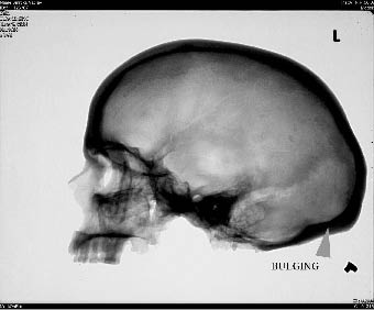
Figure 6. X-ray picture of cranium: arrow marks the bulging of the occipital bone (X-ray made by staff of the Radiodiagnostic Department of the 1st Faculty of Medicine, Charles University, Prague).

Figure 7. Three aspects of the deformed skull (photo by Anna Pankowská).
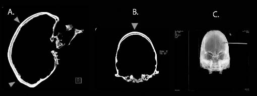
Figure 8. CT image of the cranium from three aspects: A) arrows point to the coronal suture and lambdoid suture; B) arrow points to the absence of sagittal suture; C) arrows mark the narrowing of the calvaria (scale: 4 cm) (CT image made by workers of the Radiodiagnostic department of the First Faculty of Medicine of the Charles University in Prague).

Figure 9. Comparison of two skulls: bone dissection No. 2191 and the skull from Náměšť na Hané. In left upper corner, red arrows point to the bregmatic and obelionic arachnoid granulations in the image of a plaster cast of the brain (photo by Anna Pankowská).
The acquired lesions were identified in the postcranial skeleton. At the distal ends of the tibiae lateral sclerotic layers, or Harris lines (HL) (Figure 10), are clearly visible. At the macroscopic level the tibiae are slightly curved in a latero-medial direction at their distal ends. More layers were recorded in the right tibia than in the left. At the distal ends the bone mineral density is lower than normal (Figure 8). This could be connected with sclerotic layers which are distributed from the centre of the distal part of the diaphysis to the epiphyseal line. These layers may persist from childhood into adulthood and the new transverse lines may have developed later in life. Hummert and Van Gerven (1985) argue that Harris Lines do not endure more than 10 years and disappear over time in connection with the remodeling of living bone. In our case however, they are clearly visible, particularly on right tibia.
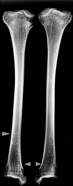
Figure 10. X-ray picture: arrows show transverse sclerotic layers in the metaphysial parts of tibiae (Harris lines) (X-ray made by staff of the Radiodiagnostic Department of the 1st Faculty of Medicine, Charles University, Prague).
5. Diagnosis
The female interred in the borrow pit at Náměšť na Hané was affected by a congenital anomaly of the skull – scaphocephaly. Scaphocephaly is a type of Craniosynostosis – CS (premature closure of cranial sutures). Craniosynostosis may or may not result in deformation of the skull (craniostenosis). Craniosynostosis is the process of premature sutural fusion, craniostenosis is the result (Cohen 1980).
This scaphocephalic skull is also affected by excessive growth of the lambdoid suture (probably bathrocephaly) which is reflected in the bulging of the occipital area. Bathrocephaly is a common developmental anomaly, resolves spontaneously, and is of no clinical significance.

Figure 11. Scheme of different types of craniosynostoses (taken from Cohen, Maclean 2000).
CS arrests calvarial expansion perpendicular to the affected suture(s). As a result, the skull will gradually assume an abnormal shape. The longer this condition exists the more profound the deformation (Oostra et al. 2005). Scaphocephaly limits the growth of the skull in a transverse direction causing an abnormally long and narrow skull (Aufderheide, Rodriguez-Martin 1997). The shape of the skull depends on which suture is defective (Figure 11).
Bowing of the tibiae could be interpreted as residual rickets or osteomalacia, but there is a range of differential diagnoses of bowing deformities, such as congenital bowing, Paget’s disease and trauma. Harris growth arrest lines were identified at the distal ends of the tibiae. There are many possible origins for these layers and the precise etiology is controversial. Most often HL’s are described in association with malnutrition, childhood infection diseases, trauma, physiological stress and generally poor living conditions, and in adults with osteoporosis (Ameen et al. 2005, Grolleau-Raoux et al. 1997, Hummert, Van Gerven 1985). A connection with osteoporosis or osteopenia could be the case in this skeleton, as the radiograph shows lower bone mineral density especially in distal ends of the tibiae. Ameen et al. (2005) state that HL may develop later in life, independent of childhood, and a new clinical study describes these layers in association with genua valga.
These two defects (premature skull suture closure and bowing of the tibiae resulting from metabolic disease) could be linked (see the study of Wang et al., 2007). These authors present the case of a preterm 6-month-old American infant who developed CS secondary to rickets. This infant developed rickets and macrocephaly by the age of 6 months. His head continued to enlarge and a CT scan obtained when the child was 2 years old revealed metopic and bilateral coronal craniosynostosis. This CT suggested increased intracranial pressure and therefore corrective cranial vault reconstruction was performed. Rickets (vitamin D deficiency) in children is associated with hypotonia, growth retardation, muscle weakness, delayed motor development, lack of sleep, etc. Consequently, vitamin D deficiency can cause the softening of cranial sutures and delaying fontanel growth, which may evoke premature fusion of cranial sutures (Wang et al. 2007).
6. Cranial sutures and classification of craniosynostoses
Craniosynostosis (premature fusion of cranial sutures) is one of the most common craniofacial anomalies with a reported incidence of up to one per 2500 live births. Males are more affected than females (Alden et al. 1999; Lajeunie et al. 1996). The most common CS is scaphocephaly, followed by brachycephaly, trigonocephaly, and a very uncommon condition caused by the fusion of the sutura lambdoidea or sutura squamosa. Cranial sutures are dense fibrous connections between cranial bones which permit a tiny amount of movement. Sutures have an important function as they are necessary for an infant’s brain and skull growth and development. Their structure and shape depends on their function and the mechanical strain they have to endure (Hajniš, Novák 1984). During childbirth the flexibility of the fibers allows the bones to overlap so that the head can pass through the birth canal without pressing on and damaging the infant’s brain. During infancy and childhood the fibers remain flexible. This allows the brain to grow quickly and protects it from minor impacts to the head – similar to shock absorbers (Cohen 2005). The physiological process of suture fusion appears after the second decade and is highly variable throughout the life (e. g. Cobb 1955, Masset 1989, Hajniš, Novák 1984). Overgrowth of fibroblast normally prevents premature fusion of cranial sutures. If this mechanism is somehow disturbed premature fusion is established. Craniosynostosis may be designated as primary or secondary. Primary craniosynostosis is the most common type of CS and examples have already been given above under simple and compound craniosynostoses. It is of unclear etiology. In secondary craniosynostosis, sutural obliteration is secondary to another disorder (metabolic, hormonal, brain disorder, hematologic, intrauterine head constraint). Craniosynostosis can be either isolated or syndromic. Syndromic craniosynostosis occurs with other defects; there are more than 90 syndromes connected with CS and in isolated cases individuals have no other abnormalities (O’Brien, Sensor 2004, Cohen 1980).
Classification of CS can be accomplished by morphology, etiology or molecular biology, since CS is pathogenetically, etiologically and phenotypically heterogeneous (Cohen 1980, 2005). Different etiology and pathogenesis can result from the same phenotype manifestation and classification of craniosynostosis is extremely difficult. The simplest categorization is the division into isolated (non – syndromic) and syndromic (Cohen 2005).
Serious forms of craniosynostosis are currently resolved surgically, preferably within the 1st month in order to prevent intracranial pressure and consequent neurological and neuropsychological disorders (Hoza et al. 2006). Hoza et al. investigated eleven children after surgery on a synostosis of the sutura sagittalis. One suffered from significant mental and motor retardation, three suffered low retardation, and the others were healthy (Hoza et al. 2006; Virtanen et al. 1999). Scaphocephaly is not usually associated with mental or physical disorders. But the greater is the deformation the greater is the chance of intracranial pressure and compression of the brain (Arnaud et al. 1995).
7. Archaeological occurrence of craniosynostosis
The Broumov ossuary in the Czech Republic contains a well investigated collection of craniosynostoses. In this collection the presence of craniosynostosis is 4.83% (3.4% in adults and 12.24% in children; Pospíšilová, Procházková, Serbouti 2003). This is much higher than modern populations were the rate is about 0.5%. The researchers explain that this is due to the fact that the entire range of craniosynostoses is currently escaping identification because it does not leave any traces of skull deformation. The difference in occurrence between adults and juveniles is explained by the fact that certain amount of plagiocephaly adjusts itself during one’s life (Pospíšilová, Procházková, Serbouti 2003). Other findings from the Czech Republic are usually deposited in anatomical collections as pathological bone dissections obtained mostly from the maceration of cadavers in the 19th century (e. g. the collection of the 2nd Pathological-Anatomical Institute of the Faculty of Medicine, Charles University, Prague). In these collections we find cases of scaphocephaly and others kinds of craniosynostoses, some with clinical records and plaster brain casts (plaster brain casts are deposited in the 2nd Pathological-Anatomical Institute of the Faculty of Medicine, Charles University, Prague).
Oostra et al. (2005) analyzed the skulls with CS and suture-related conditions in the extraordinary collection of the Vrolik Museum in Amsterdam. The osteological collection from Vrolik has existed since the 19th century and comprises more than 5,000 specimens of human and nonhuman anatomy, embryology and pathology. On external examination they found 58 skulls with craniosynostosis, out of which 26 were scaphocephalic (Oostra et al. 2005).
Other publications describe findings of CS from the USA (Hohenthal, Brooks 1960, Bennet 1967, Duncan, Stojanowski 2008). One case of craniosynostosis (plagiocephaly) from an archaeological site in Georgia (USA) was used as a tool for positive identification of human remains (Duncan, Stojanowski 2008). Gracia et al. (2009) reported the oldest case of craniosynostosis (lambdoid single suture synostosis) on a Middle Pleistocene immature human specimen at Sima de los Huesos. According to the authors, this defect occurred before birth and influenced the individual’s motor-cognitive skills (Gracia et al. 2009)
There are two rare cases of craniosynostosis (scaphocephaly) dated to the Eneolithic. Both were recorded at archaeological sites in Spain, at Cova del Palanques in Valencia, and Navarres (Campillo 1993 in Aufderhide, Rodriguez-Martin 1998). Other scaphocephalic skulls are known from the Guanches population of the Canary Islands (Spitery 1983 in Aufderhide, Rodriguez-Martin 1998), 20–25th Dynasty Egypt, the Karga site (Ortner 2003), and from Cinco Cerros in Peru (Ortner 2003).
8. Summary and conclusion
During the rescue excavation in Náměšť na Hané a settlement of the Funnel Beaker culture was investigated. In feature 7 a female skeleton was found interred, in a partially filled borrow pit. At the time of the burial this borrow pit probably did not serve its original function. The well preserved skeleton could be radiocarbon dated, and this was carried out at the University of Georgia in the USA (UGMAS # 05712). The uncalibrated date is 4590±30 BP (3380–3330 BC cal. ). This date corresponds to the late phase of the Funnel Beaker culture or to the end of the Boleraz horizon and the beginning of the classical phase of the Baden culture. However, pottery goods from pit No. 7 and other features are dated to the Early Funnel Beaker culture. The situation may be explained as a later inhumation at an abandoned settlement pit.
The woman suffered from a congenital anomaly of the skull – scaphocephaly – and from non-specific diseases which appeared in the form of Harris lines on the tibiae. This pathological finding is important in association with the archaeological context and the circumstances of the funeral and may be evidence of the treatment of defective individuals at that time. While this woman may have not been affected by a mental disorder her appearance differed markedly from what was normal in the surrounding society. It is possible that the funeral practice was a reflection of her social status. From an anthropological point of view this is a uniquely well-preserved find from the period, and is of local and worldwide importance.
Acknowledgements
The authors would like to thank RNDr. Vítezslav Kuželka for providing bone dissections from the collection of the 2nd Pathological Institute of the National Museum in Prague. Furthermore, we thank workers of the Radiodiagnostic Department of the 1st Faculty of Medicine, Charles University, Prague, MUDr. Josef Horejš for consultations, and RNDr. Miriam Nývltová–Fišáková, Ph. D., for analysis of the tiny beads of freshwater pearl mussel shells.
References
ALDEN, T. D., LIN, K. Y., JANE, J. A. 1999: Mechanisms of premature closure of cranial sutures. Child’s Nervous System 15, 670–675.
AMEEN, S., STAUB, L., ULRICH, S., VOCK, P., BALLMER, F., ANDERSON, S. E. 2005: Harris lines of the tibia across centuries: a comparison of two populations, medieval and contemporary in Central Europe. Skeletal Radiology 34, 279–284.
ARNAUD, E., RENIER, D., MARCHAC, D. 1995: Prognosis for mental function in scaphocephaly. Journal of Neurosurgery 83, 476–479.
AUFDERHEIDE, A. C., RODRÍGUEZ-MARTÍN, C. 1998: The Cambridge Encyclopedia of Human Paleopathology. Cambridge University Press.
BEDNARCZYK, J., CZERNIAK, L., KOŚKO, A. 1980: Z badań nad zespołem osadniczym ludności k kręgu kulturi ceramiki wstęgowej w Kruszy Zamkowej (woj. bydgoskie) stan. 3 (część sepulkralna), Sprawozdania Archaeologiczne XXXII, 55–83.
BANNER, J. 1956: Die Péceler Kultur. Budapest.
BÁTORA, J. 1982: Ekonomicko-sociálny vývoj východného Slovenska v staršej dobe bronzovej, Slovenská Archeológia XXX, 249–314.
BÁTORA, J. 2000: Das Gräberfeld von Jelšovce/Slowakei. Ein Beitrag zur Frühbronzezeit im nordwestlichen Karpatenbecken. Prähistorische Archäologie in Südosteuropa Band 16, Kiel.
BÁTORA, J. 2002: Contribution to the Problem of “Craftsmen” Graves at the End of Eneolithic and ih the Early Bronze Age in Central, Western and Eastern Europe. Slovesnká Archeológia L, 179–228.
BENNET, K. A. 1967: Craniostenosis: A review of etiology and a report of new cases. American Journal of Physical Anthropology 27, 1–10.
BIALEKOVÁ, D. 2000: Hrob skrčenca z Pobedima. In: ČECH, P. – DOBEŠ, M. (eds. ), Sborník Miroslavu Buchvaldkovi, Most, 21–26.
BROOKS, S. T, SUCHEY, J. M. 1990: Skeletal Age Determination Based on pubis: A Comparison of the Acsadi-Nemeskeri and Suchey-Brooks Methods. Human Evolution 5, 227–238 (in White TD. 2000. Human osteology. 2nd Ed. New York: Academic Press).
BUCHVALDEK, M. 1986: Kultura se šňůrovou keramikou ve střední Evropě. I. Skupiny mezi Harcem a Bílými Karpaty. Praehistorica XII. Praha.
CAMPILLO, D. 1993: Paleopatología: los primeros vestigios de la enfermedad (Paleopathology. The first evidence of disease). Segunda Parte. Barcelona: Fundación Uriach 1838.
CHLEBORÁD, M. 1934: Pravěké hroby durinských skrčků na Bučovsku a v okolí. Zvl. otisk z ročenky spořitelny města Bučovic za rok 1934. Bučovice 1934.
COBB, W. M. 1955: The Age Incidence of Suture Closure. American Journal of Physical Anthropology 13, 2, 394–401.
COHEN, M. M. 1980: Perspectives on Craniosynostosis. The Western Journal of Medical Genetics 132, 507–513.
COHEN, M. M. 2005: Editorial perspectives on Craniosynostosis. American Journal of Medical Genetics 136A, 313–326.
COHEN, M. M., MacLEAN, R. E. 2000: Craniosynostosis: diagnosis, evaluation, and management, 2nd ed. New York: Oxford University Press.
CZEKAJ-ZASTAWNY, A. 2009: Obrządek pogrzebowy kultury ceramiky wstęgowej rytej. In: Czekaj-Zastawny, A. (ed. ): Obrządek pogrzebowy kultur pochodzienia naddunajskiego w nwolicie Polski południowo-wschodniej (5600/5500–2900 BC). The Funerary rite of the Danubian cultures in the Neolithic of southeastern Poland (5600/5500–2900 BC), Kraków, 25–51.
DOBEŠ, M. 1998: Gräber der Kugelamphorenkultur in Nordwestböhmen. Saarbrücker Studien und Materialien zur Altertumskunde 6/7, 133–179.
DOBEŠ, M. 2008: Kultura kulovitých amfor. In: Neústupný, E. (ed. ), Archeologie pravěkých Čech/4. Eneolit. Praha, 115–122.
DOBEŠ, M., Buchvaldek, M. 1993: Katalog šňůrové keramiky v Čechách VIII. Mostecko. Praehistorica XX. Praha, 197–258.
DROZDOVÁ, E. 2004: Základy osteometrie. Panoráma biologické a sociokulturní antropologie. Modulové učební texty pro studenty antropologie a “příbuzných” oborů. Nadace Universitas Masarykiana v Brně.
DUNCAN, W. N., STOJANOWSKI, C. M. 2008: A Case of Squamosal Craniosynostosis from the 16th century Southeastern United States. International Journal of Osteoarchaeology 18, 407–420.
GIRIĆ, M. 1971: Mokrin. Nekropola ranog bronzanog doba. Dissertationes et Monographie T. XI, Beograd 1971.
GRACIA, A., ARSUAGA, J. L., MARTÍNEZ, I., LORENZO, C., CARRETERO, J. M., BERMÚDEZ de CASTRO, J. M., CARBONELL, E. 2009: Craniosynostosis in the Middle Pleistocene human Cranium 14 from the Sima de los Huesos, Atapuerca, Spain. Proceedings of the National Academy of Sciences 16, 6573–6578.
GROLLEAU-RAOUX, J., CRUBÉZY, E., ROUGE, D., BRUNGE, J., SAUNDERS, S. P. 1997: Harris lines: A study of age-associated bias in counting and interpretation. American Journal of Physical Anthropology 103, 209–217.
GRYGIEL, R. 2004: Neolit i początky epoki brązu w rejonie Brześcia Kujawskiego i Osłonek, Tom I Wczesny neolit. Kultura ceramili wstęgowej rytej – The Neolithic and Early Bronze Age in the Brześć Kujawski and Osłonki Region, Volume I Early Neolithic Linear Pottery Culture. Łódź.
GRYGIEL, R. 2004: Neolit i początky epoki brązu w rejonie Brześcia Kujawskiego i Osłonek, Tom II/Część 1–3 Środkowy neolit. Grupa Brzesko-Kujawska kultury lendzielskiej – The Neolithic and Early Bronze Age in the Brześć’Kujawski and Osłonku Region, Volume II/Part 1–3. Middle Neolithic The Brześć Kujawski Group of the Lengyel Culture. Łódź.
HAJNIŠ, K., NOVÁK, J. T. 1984: Srůst švů lebeční klenby. Avicenum. Praha.
HAVEL, J. 1981: Hrob kultury se šňůrovou keramikou v Praze 8-Čimicích, Praehistorica VIII – Varia Archaeologica 2, 67–71.
HOHENTHAL, W. D., BROOKS, S. T. 1960: An Archaeological Scaphocephal from California. American Journal of Physical Anthropology 18, 59–67.
HORŇANSKÝ, J., SKUTIL, J. 1950: Hromadný hrob kultury s keramikou malovanou vr Džbánicích u Moravského Krumlova, Obzor prehistorický XIV, 333–336.
HOZA, D., ŽÁČKOVÁ, J., HOŘÍNEK, D. 2006: Výsledky neuropsychologických vyšetření vývojovými škálami Bayleyové u dětí po operaci skafocefalie. Česká a slovenská neurologie a neuropsychologie. Časopis českých a slovenských neurologů a neurochirurgů. Česká lékařská společnost J. E. Purkyně. Supplementum 3.
HUMMERT J. R., Van GERVEN, D. P. 1985: Observation on the formation and the persistence of radipaque transverse lines. American Journal of Physical Anthropology 66, 297–306.
KADROW, S. 1998: Osteuropäische Beziehungen des epischnurkeramischen karpatenländischen Kulturkreises in der Frühbronzezeit. In: Hänsel, B., Machnik, J. (eds. ): Das Karpatenbecken und die osteuropäische Steppe. Nomadenbewegungen und Kulturaustausch in den vorchristlichen Metallzeiten (4000–500 v. Chr. ), Südosteuropa-Schriften 20, Prähistorische Archäologie in Südosteuropa 12. München – Rahden/Westf., 253–260.
KADROW, S., MACHNIK, A., MACHNIK, J. 1992: Iwanowice, stanowisko babia Góra. Część’II. Cmentarzysko z wczesnego okresu epoki brązu. Kraków.
KLČO, M., KRUPA, V. 2008: Hrob kultúry so šňúrovou keramikou z Krakovian, Hlas Krakovian roč. 6, č. 2, 7.
KNUSSMAN, R. 1988: Anthropologie, Handbuch der vergleichenden Biologie des Menschen (4. Auflage des Lehrbuchs der Anthropologie begründet von Rudolf Martin), Band I und II. Jena, New York, Stuttgart: Gustav Fisher.
KRUMPEL, J. 2009: Vier Gräber der Badener Kultur aus Ratzersdorf, Niederösterreich. Eine Neubewertung der Bestattungssitten der Badener Kultur in ihrer österreichischen Verbreitung. Fundberichte aus Österreich 47, 2008, Wien, 99–150.
LAJEUNIE, E., MERRER, M. L., BONAÏTI-PELLIE, MARCHAC, D., RENIER, D. 1996: Genetic Study of Scaphocephaly. American Journal of Medical Genetics 62, 282–285.
LOVEJOY, C. O., MEINDL, R. S., PRZYBECK, T. R. 1985: Chronological Metamorphosis of the auricular Surface of Illium: A New Method for the Determination of Adult Skeletal age at Death. American Journal of Physical Anthropology 68:15–28. (take from White TD. 2000. Human osteology. 2nd Ed. New York: Academic Press).
MACHNIK, J. 1979: Krąg kulturowy ceramiki sznurowej. In: Godlowska, M., Kulczyska-Leciejewiczowa, A., Machnik, J., Wislanski, T. (Eds. ): Prahistoria ziem Polskich, Tom II Neolit. Wrocław – Warszawa – Kraków – Gdańsk, 337–411.
MASSET C. 1989: Age estimation on the basis of cranial sutures. In Iscan, M. Y. (Ed. ): Age Markers in the Human Skeleton. Springfield: Charles C Thomas, 71–103.
MARKEVIČ, V. I. 1981: Pozdnetripo’lskije plemena severnoj Moldavii. Kišinev.
MEDUNOVÁ-BENEŠOVÁ, A. 1964: Eneolitické výšinné sídliště Staré Zámky v Brně – Líšni. Památky archeologické LV, 91–155.
MOUCHA, V. 1958: Příspěvek k časovému zařazení eneolitických zápon. Archeologické rozhledy X, 40–44, 62–78.
MOUCHA, V. 1992: Die Schnurkeramik und die Glockenbecherkultur in Böhmen. In: Die kontinentaleuropäischen Gruppen der Kultur mit Schnurkeramik. Schnurkeramik-Symposium Štiřín 1990. Praehistorica XIX, Praha, 81–87.
MURAIL, P., BRUZEK, J., HOUET, F., CUNHA, E. 2005: DSP: a tool for probabilistic sex diagnosis using worldwide variability in hip bone measurements. Bulletin set Memoires de la Societe d’Anthropologie de Paris, n. s., t. 17, 3–4: 167–176.
NEUSTUPNÝ, E. 2008: Kultura se šňůrovou keramikou. In: Neustupný, E. (Ed. ), Archeologie pravěkých Čech/4. Eneolit. Praha, 124–147.
NEUSTUPNÝ, E., SMRŽ, Z. 1989: Čachovice – pohřebiště kultury se šňůrovou keramikou a zvoncovitých pohárů, Památky archeologické LXXX, 232–383.
O’BRIEN, T. G., SENSOR, K. P. 2004: On the classification of abnormal head shape: Interpreting artificial cranial deformation and craniosynostosis. American Journal of Physical Anthropology Supplement 40, 159–167.
OOSTRA, R. J., WOLK, S., MAAS, M., HENNEKAM, C. M. 2005: Malformations of the axial skeleton in the museum Vrolik: II: Craniosynostoses and suture-related conditions. American Journal of Medical Genetics 136A, 327–342.
ORTNER, J., 2003: Identification of Pathological Conditions in human skeletal remains. 2nd edition. Academic Press. San Diego.
PAVELČÍK, J. 1992: Příspěvek k absolutnímu datování osady lidu s kanelovanou keramikou u Lipníka nad Bečvou, Časopis Slezského zemského muzea série B, 41, 193–195
PAVELČÍK, J. 1993: Lid s kanelovanou keramikou. In: Podborský, V. (Ed. ): Pravěké dějiny Moravy. Brno, 179–190.
PAVELČÍK, J. 2002: Neolithikum und Äneolithikum in Nordmähren und Schlesien (Troppauer Gebiet) im Lichte der 14C-Daten, Preistoria Alpina 37, 2001, Trento, 333–336.
PEARSON, K., 1935: Note on section of Dr. K. Wagners memoir. Biometrika 17:53–56.
PODBORSKÝ, V. 1993: Lid s moravskou malovanou keramikou. In: Podborský, V. (Ed. ): Pravěké dějiny Moravy. Brno, 108–150.
POSPÍŠILOVÁ, B., PROCHÁZKOVÁ, O., SERBOUTI K. 2003: Paleopatologické nálezy předčasné obliterace věnčitého švu. Acta Medica (Hradec Králové) SUPPL 46, 13–21.
ŠEBELA, L. 1999: The Corded Ware Culture in Moravia and the adjacent Part of Silesia (Catalogue), Fontes Archaeologiae Moravicae XXIII. Brno.
SJØVOLD, T. 1990: Estimation of stature from long bones utilizing the line of organic correlation. Human Evolution 5, 431–447.
ŠMÍD, M. 2006: Pohřebiště nitranské kultury ze Slatinic, okres Olomouc, Slovenská Archeológia LIV, 1–32. Adress: Tetín 8, Prostějov, announced on 16th September 2009.
SPITERY, E. 1983: La Paléontologie des maladies osseuses constitutionelles (The Paleontology of Constitutional Bone Diseases). Paris: Editions du CNRS.
STADLER, P., DRAXELER, S., FRIESINGER, H., KUTSCHERA, W., PRILLER, A., ROM, W., STEIER, P., WILD, E. M. 2001: Absolute Chronology for Early Civilizations in Austria and Central Europe using 14C Dating with Accelerator Mass Spectrometry with special Results for the Absolute Chronology of the Baden Culture. In: Cernovodă III – Boleráz. Ein vorgeschichtliches Phänomen zwischen dem Oberrhein und der unteren Donau, Symposium Mangalia/Neptun (18. –24. Oktober 1999), Studia Danubiana series Symposia II, Bucureşti, 541–562.
STOJANOWSKI, C. M. SEIDEMANN, R. M., DORAN, G. H. 2002: Differential Skeletal Preservation at Windover Pond: Causes and Consequences. American Journal of Physical Anthropology 119, 15–26.
TICHÝ, R. (Ed. ) in press: Hroby, pohřby a lidské pozůstatky na pravěkých a středověkých sídlištích. Supplementum 2, Živá archeologie.
TOČÍK, A. 1978: Záchranný výskum v Komjaticiach, Archeologické výskumy a nálezy na Slovensku v roku 1977, Nitra, 246–272.
TOČÍK, A. 1979: Výčapy-Opatovce a ďalšie pohrebiská zo staršej doby bronzovej na juhozápadnom Slovensku. Materialia Archaeologica Slovaca 1. Nitra.
VIRTANEN, R., KORHANEN, T., FAGERHOLM, J., VILJANTO, J. 1999: Neurocognitive Sequelae of Scaphocephaly. Pediatrics 103, 791–795.
VRÁNA, J. 2010: Nálezová zpráva ze záchranného archeologického výzkumu, Náměšť na Hané, Excavation report nr. 252/2010. Deposited: Archaeological Centre Olomouc.
VRÁNA, J., PANKOWSKÁ, A. 2010: Kostrový pohřeb v sídlištním objektu v Náměšti na Hané. In: Bém, M., Peška, J. (Eds. ): Ročenka 2009, Olomouc, 70–93.
WANG, P. I., MARCUS, J. R., HERBERT, E. F., MUKUNDAN, S. 2007: Craniosynostosis secondary to rickets: manifestations on computed tomography. Radiology Case Reports 2, 43–54.
WIŚLAŃSKI, T. 1979: Plemiona kultury amfor kulistych. In: Henesel, W, Wiślański, T. (Eds. ): Neolit (Prahistoria ziem Polskich, t. II). Wrocław – Warszawa – Kraków – Gdańsk, 261–299.
WŁODARCZAK, P. 2006: Kultura ceramiki sznurowej na Wyżynie Małopolskiej. PAN: Kraków.
ZAKOSCIELNA, A. 2009: Obrządek pogrzebowy kultury Lubelsko-wołyńskiej. In: Czekaj-Zastawny, A. (Ed. ): Obrządek pogrzebowy kultur pochodzienia naddunajskiego w nwolicie Polski południowo-wschodniej (5600/5500–2900 BC). The Funerary rite of the Danubian cultures in the Neolithic of southeastern Poland (5600/5500–2900 BC). Kraków, 107–154.
ZÁPOTOCKÝ, M. 2008: Kultura nálevkovitých pohárů ve starším eneolitu. In: E. Neustupný, (Ed. ): Archeologie pravěkých Čech/4. Eneolit. Praha, 61–82.